

Movement Analysis
Detailed analysis of movement is a complex activity requiring sophisticated equipment. However, the fundamental analysis of motion can be done visually and should involve the following:
- A description of the actual actions which occur at the joints involved
- The plane(s) in which the movement occurs
- The muscles producing the movement
- The function of the muscles involved (agonists, antagonists, synergists & fixators)
- The type of contraction (isotonic - concentric or eccentric, isometric)
- The range of the muscle action (inner, middle, outer)
Analysis of Sprinting
The running leg action occurs in a sagittal plane about a frontal axis and involves the hip, knee and ankle joints.
The hip's bones are the femur and pelvic girdle, which form a ball and socket joint.
The knee bones involved are the femur and tibia, which form a hinge joint.
The ankle bones are the tibia and calcaneus, which form a modified joint.
Each of these joints produces two actions, one when the leg is in contact with the ground (driving phase) and one when the leg is not in contact with the ground (recovery phase).
Driving Phase
| Joints involved | Action | Agonist Muscle |
| Hip | Extension & hyperextension | Gluteal muscles (gluteus maximus and gluteus minimus) and Hamstrings (biceps femoris, semimembranosus, semitendinosus) |
| Knee | Extension | Quadriceps group of muscles (rectus femoris, vastus medialis, vastus lateralis and vastus intermedialis) |
| Ankle | Plantar flexion | Gastrocnemius |
Recovery phase
| Joints involved | Action | Agonist Muscle |
| Hip | Flexion | Iliopsoas |
| Knee | Flexion | Hamstrings (biceps femoris, semimembranosus, semitendinosus) |
| Ankle | Dorsiflexion | Tibialis anterior |
Analysis of Throwing
Throwing comprises of two phases, the preparatory phase and the throwing phase. Most actions are rotational in the transverse plane and longitudinal axis and the two joints primarily involved are the elbow and shoulder. The elbow is a hinge joint formed by the humerus and ulna. The shoulder is a ball and socket joint formed between the humerus and the scapula. Note: The javelin is incorrectly held. See the Javelin section for more details. |
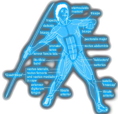 |
Preparatory phase
| Joints involved | Articulating bones | Action | Agonist Muscle |
| Shoulder | Humerus & scapula | Horizontal hyperextension | Posterior deltoids and latissimus dorsi |
| Elbow | Humerus & ulna | Extension | Triceps brachii |
Throwing phase
| Joints involved | Articulating bones | Action | Agonist Muscle |
| Shoulder | Humerus & scapula | Horizontal flexion | Anterior deltoids and Pectoralis major |
| Elbow | Humerus & ulna | Flexion | Biceps brachii |
Analysis of racket strokes
There are two phases to striking a ball with a racket, the preparatory phase and the striking phase. Most actions are rotational in the transverse plane, and longitudinal axis and the three joints concerned are the wrist, elbow and shoulder. The elbow is a hinge joint formed by the humerus and ulna. The shoulder is a ball and socket joint formed between the humerus and the scapula. The wrist forms a condyloid joint between the ulna and carpal bones. |
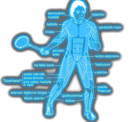 |
Preparatory Phase
| Joints involved | Articulating bones | Action | Agonist Muscle |
| Wrist | Ulna & carpal Radius &ulna |
Supination | Supinator |
| Elbow | Humerus & ulna | Extension | Triceps brachii |
| Shoulder | Humerus & scapula | Horizontal hyperextension | Posterior deltoid and latissimus dorsi |
Striking Phase
| Joints involved | Articulating bones | Action | Agonist Muscle |
| Wrist | Ulna & carpal Radius & ulna |
Pronation | Pronator teres |
| Elbow | Humerus & ulna | Flexion | Biceps brachii |
| Shoulder | Humerus & scapula | Horizontal flexion | Pectoralis major and Anterior deltoid |
| Trunk | Rotation | External obliques |
Analysis of jumping
The action in jumping takes place in a sagittal plane about a transverse axis and involves the hip, knee and ankle joints. The hip's bones are the femur and pelvic girdle, which form a ball and socket joint. The bones of the knee involved are the femur and tibia which form a hinge joint. The bones of the ankle involved are the tibia and calcaneus which form a modified joint. |
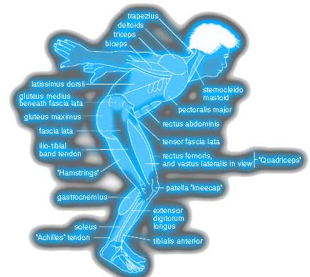 |
| Joints involved | Action | Agonist Muscle |
| Hip | Extension &hyperextension | Gluteal muscles (gluteus maximus and gluteus minimus) and Hamstrings (biceps femoris, semimembranosus, semitendinosus) |
| Knee | Extension | Quadriceps group of muscles (rectus femoris, vastus medialis, vastus lateralis and vastus intermedialis) |
| Ankle | Plantar flexion | Gastrocnemius |
Analysis of Kicking
The kicking action takes place in a sagittal plane about a frontal axis and involves the hip, knee and ankle joints. The hip's bones are the femur and pelvic girdle, which form a ball and socket joint. The bones of the knee involved are the femur and tibia which form a hinge joint. The bones of the ankle involved are the tibia and calcaneus which form a modified joint. Kicking comprises of two phases, the preparatory phase and the kicking phase. |
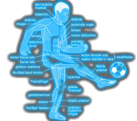 |
Preparatory Phase
| Joints involved | Action | Agonist Muscle |
| Hip | Extension & hyperextension | Gluteal muscles (gluteus maximus and gluteus minimus) |
| Knee | Flexion | Hamstrings (biceps femoris, semimembranosus, semitendinosus) |
| Ankle | Plantar flexion | Gastrocnemius |
Kicking phase
| Joints involved | Action | Agonist Muscle |
| Hip | Flexion | Iliopsoas |
| Knee | Extension | Quadriceps group of muscles (rectus femoris, vastus medialis, vastus lateralis and vastus intermedialis) |
| Ankle | Plantar flexion | Gastrocnemius |
Agonist, Antagonist, Fixator & Synergist Muscles
A question often asked is to identify the Agonist, Antagonist, Fixator & Synergist Muscles. When kicking the ball then:
- Agonist - Quadricep muscles
- Antagonist - Hamstring muscles
- Fixator - Gluteus Maximus
- Synergist - Abdominal muscles
Page Reference
If you quote information from this page in your work, then the reference for this page is:
- MACKENZIE, B. (2007) Movement Analysis [WWW] Available from: https://www.brianmac.co.uk/moveanal.htm [Accessed